

Mole Removal in Frankfurt
Moles can change over the course of a person’s life or be perceived as bothersome in everyday life. At our private practice for dermatology and skin surgery in Frankfurt, we evaluate your moles from a dermatological perspective and then remove them on an outpatient basis. Depending on the findings, we may opt for traditional surgical removal for laboratory analysis or for ablative laser procedures.
What types of moles are there?
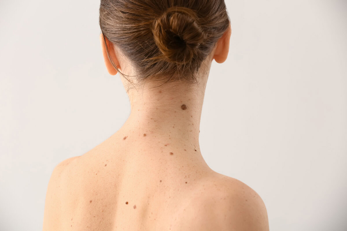
Almost everyone has them: moles (nevi). They are benign skin lesions caused by a cluster of pigment-producing cells (melanocytes) or blood vessels. The average adult has between 10 and 40 of these spots.
But not all moles are the same. They differ in color, shape, depth, and origin.
Classic Pigmented Lesions (Melanocytic Nevi)
These moles are caused by the proliferation of melanocytes, the cells responsible for our skin’s tanning. Depending on which layer of the skin the cells are located in, there are three main types:
Junctional nevus:
- Appearance: Flat, sharply defined, usually dark brown to black.
- Location: Pigment cells are located in the outermost layer of the epidermis.
Compound nevus (mixed type):
- Appearance: Slightly raised, usually an even brown color, sometimes with a slightly rough surface.
- Location: The cells are located in both the upper dermis and the underlying dermis.
Dermal nevus (true birthmark):
- Appearance: Distinctly raised, knot- or button-like, often skin-colored to light brown. Small hairs often grow from them.
- Location: The pigment cells are located deep within the dermis.
Special and Atypical Forms
Some moles differ from the typical appearance. They often require a little more attention:
Atypical / Dysplastic Nevus:
- These lesions are usually larger than 5 mm, irregular in shape, and multicolored (a mixture of brown, red, and pink hues). Although they are benign, they are considered an indicator of an increased risk of skin cancer and should be checked regularly by a doctor.
Halo nevus (Sutton nevus):
- A brown birthmark surrounded by a white, depigmented ring. In this case, the body’s own immune system is trying to break down the pigment cells. The birthmark often fades on its own over time.
Blue nevus:
- A small, firm nodule that is blue-gray or blue-black in color. The unusual color is caused by the deep location of the pigment cells in the skin, which refract light differently.
Congenital nevus (birthmark):
- These birthmarks are present at birth or develop shortly thereafter. They can range from tiny spots to large, hairy areas.
Vascular Birthmarks
In colloquial language, skin lesions that do not consist of pigment cells at all, but rather of blood vessels, are also referred to as birthmarks:
- Stork bite (fire mark): Pale red spots on the back of the head, forehead, or eyelids of newborns, which usually fade during the first year of life.
- Hemangioma (“blood blister”): Benign growths of blood vessels that appear as small, bright red spots or raised areas.
When should you see a dermatologist?
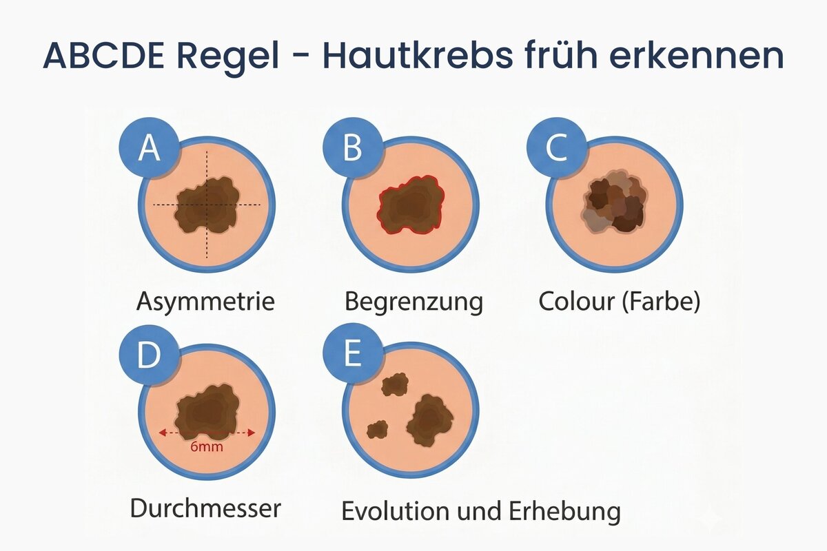
Most moles are completely harmless. However, some types of moles—or new moles that appear on the skin—can develop into malignant lesions. The simple ABCDE rule can help you check your moles yourself:
- Asymmetry: The birthmark is not a uniform circle or oval.
- Border: The edges are blurred, jagged, or frayed.
- Color: The spot has different shades (e.g., brown, black, reddish).
- Diameter: The birthmark is larger than 5 mm.
- Development: It changes in shape, size, or color, or starts to itch or bleed.
If you notice a mole that has changed or a new one that has appeared, please have it checked during your annual skin cancer screening at our practice.
When should or can you have a birthmark removed?
The reasons for removal are usually either medical or cosmetic in nature.
- Medical Indication: If a mole changes in shape, color, or size (suggesting skin cancer or a malignant mole) or if it bleeds or itches, it should be examined by a dermatologist immediately and, if necessary, surgically removed.
- Functional reasons: Protruding moles on the face, neck, or in areas where bra straps, belts, or razors rub can be bothersome or painful
Safety first: Every mole that is surgically removed at our practice is routinely sent to our partner laboratory for a detailed tissue examination (histology) to ensure that any malignant changes are ruled out.
Methods for Mole Removal at Our Frankfurt Practice
Depending on the findings, tissue depth, and location, we will select the least invasive procedure for you:
Surgical removal
- When to use: For all medically significant moles, as well as deep-seated, raised pigmented lesions.
- Procedure: Under local anesthesia, the birthmark is precisely excised with a scalpel. The wound is then closed using the most advanced and delicate suturing techniques, often with continuous sutures placed beneath the skin to minimize visible scarring.
Removal Using a Laser
- When to use: Only for moles that are clearly benign or slightly raised.
- Procedure: The pigmented tissue is precisely ablated layer by layer using high-energy laser light. The treatment usually leaves only a fine abrasion that heals quickly.
How is a mole removed?
- Initial Examination & Consultation
Using reflected-light microscopy (dermatoscopy), we will carefully evaluate your mole and work with you to determine the best treatment option. - Gentle Removal
The procedure is performed on an outpatient basis at our practice under local anesthesia. You will generally not feel any pain during the brief procedure. - Wound Care & Aftercare
We’ll provide you with an appropriate wound dressing as well as detailed tips on scar care to ensure optimal healing. - Histological examination
If the tissue is surgically removed, it is analyzed histologically. We usually have the results within a few days.
FAQ - Frequently Asked Questions After Mole Removal
What should I do during the first few days after the procedure?
Keep the wound clean and dry. Avoid strenuous physical activity or sports during the first few days to prevent strain on the stitches or wound and to avoid further bleeding.
When can I take a shower again after having a mole removed?
After the removal of a mole, the right time to take your first shower depends primarily on the type of wound dressing used. If a special waterproof bandage is used, a short shower is usually possible without any problems as early as the first or second day after the procedure; however, you should avoid full baths or prolonged contact with water for the time being. If, on the other hand, a regular bandage was applied, the wound must remain completely dry for at least 48 hours. To avoid compromising the healing process, it is recommended to gently pat the bandage dry after showering or to replace it immediately with a fresh one if moisture has come into contact with the wound.
How soon after having a mole removed can I resume exercising or swimming?
Some patience is also required when it comes to sports and wellness activities after the procedure. Light exercise, such as walking or gentle workouts, is usually recommended only after the sutures have been removed. You should wait at least three to four weeks before engaging in intense sports or strength training, especially after procedures on the back or joints, as the strong muscle tension could otherwise unnecessarily stretch or reopen the fresh scar. You should also avoid swimming, saunas, and full-body baths until the stitches have been removed and the wound is completely closed.
How can I best support scar healing?
Do not apply ointments or oils until the stitches have been removed and the wound has completely healed. Starting in the third week, massage the fresh scar several times a day using circular motions with a special scar cream or skin care oil to keep the tissue supple. Silicone patches or gels can help prevent raised scars.